Opening This Fall
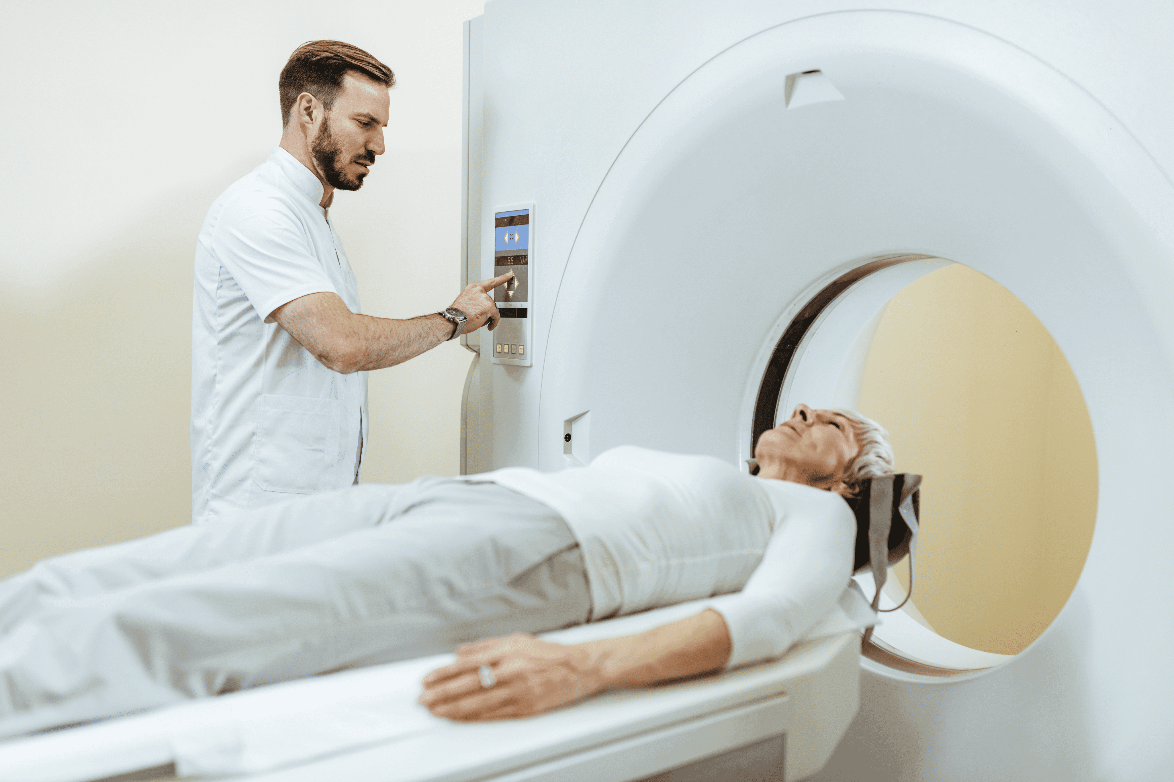
The hip is one of the body's largest and most complex joints, and diagnosing the source of hip pain is not always straightforward. A hip MRI uses a powerful magnetic field and radiofrequency pulses to generate cross-sectional images of the hip joint and its surrounding structures. It captures the muscles, tendons, cartilage, and bone in precise detail. All of this is achieved without exposing you to ionizing radiation, making it a safe diagnostic option for most patients.
When X-rays or physical examinations leave questions unanswered, hip MRI gives your physician the diagnostic clarity needed to identify what is actually happening inside the joint. For patients across the Brazos Valley searching for an MRI near me, Aggieland Imaging performs every scan on our Siemens Espree 1.5T wide-bore scanner, reviewed by a qualified radiologist who prepares a detailed report for your care team.
Hip pain has many possible causes, and pinpointing the right one requires the right imaging tool. Your physician may order a hip MRI near me when symptoms persist beyond what conservative treatment can address, or when a more precise diagnosis is needed before planning further care.
Conditions and situations that commonly lead to a hip MRI referral include:
Among athletes, hip labral tears are one of the most common findings on a sports injury MRI.
A hip MRI provides a comprehensive view of the structures that make the hip joint function. This includes the femoral head, the acetabulum, the surrounding cartilage surfaces, the hip labrum, and the major tendons and muscle groups that control hip movement.
One area where hip MRI is particularly valuable is in identifying labral pathology. A labrum tear hip MRI in Texas can reveal the precise location and extent of labral damage. This information is critical for determining whether a patient is a candidate for arthroscopic repair or whether non-surgical management is more appropriate.
Hip MRI is also the preferred imaging tool for detecting avascular necrosis at an early stage. Studies show MRI has a sensitivity of over 99% for identifying AVN, making it significantly more reliable than plain radiography for this condition. Early detection gives physicians the opportunity to intervene before irreversible bone collapse occurs.
What makes hip MRI clinically valuable: